Universal language of discovery
Jianfeng Lin’s journey from southeast Asia to a biology lab in northeast America sparks innovation
From where Jianfeng Lin was born and raised, he could gaze across the six-mile wide Taiwan Strait and make out the skyline of Taipei, the capital of the sovereign Chinese territory and island Taiwan.
His hometown, Xiamen, is a coastal metropolis of three-and-a-half million people in southeastern China. A historic seaport for foreign trade dating back to the Song Dynasty, Xiamen is where Lin developed a curiosity for the natural world. It’s where he studied microbiology as a Ph.D. student at Xiamen University.
Lin also saw what lay beyond the shore of Taipei. He wanted to further his research and begin his career as a biologist across the Pacific in America.
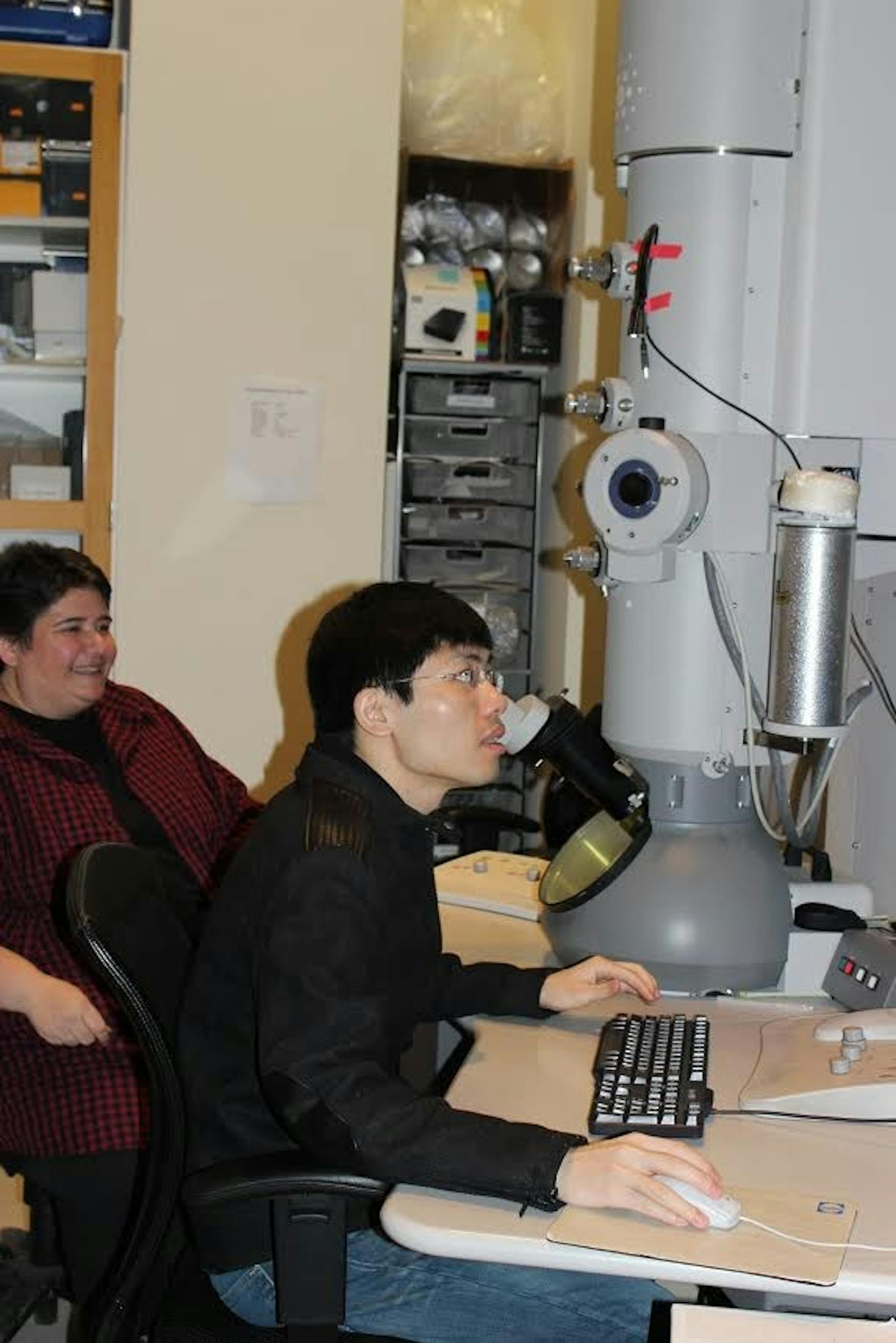
Lin is now the 33-year- old postdoctoral research assistant of Prof. Daniela Nicastro (BIOL). He was the lead scientist for two breakthrough studies last year. The most recent was published in the Dec. 4 issue of Nature Communications, a widely circulated journal that encompasses all scientific fields.
In an email to the Justice, Nicastro described Lin as “calm, respectful, at the beginning he was more reserved and I had to encourage him initially to step into a leadership role.” After nearly seven years as her postdoctoral research assistant, Nicastro recently promoted Lin to permanent senior scientist.
Lin’s journey to America is a new kind of American dream. Lin didn’t come to America to escape a war or to broaden horizons for his family. Instead, Lin came as a scientist hopeful to further his studies and career in the labs of a place that is in his own words the “most advanced and competitive country in the world.”
Lin is comfortable in his new workplace. “The U.S. is an immigrant country. I don’t have a strong feeling that I am a foreigner, and I can enjoy the diverse cultures,” he wrote in an email to the Justice.
In the summer of 2008, Lin was alerted by a friend of the opportunity in Nicastro’s lab. He would be working as a postdoctoral research assistant with a focus on cilia and flagella, a subject that he knew little about at the time.
Among the applications for the position, many from China, “Jianfeng’s application caught my eye, because he included a gorgeous image of a complex 2D electrophoresis gel,” Nicastro wrote.
Nicastro is especially fond of beautiful images, as her lab specializes in high-resolution imaging of a variety of cell bodies, including cilia and flagella.
The team’s most recent breakthrough study involves the development of a technique that allows scientists to study the building blocks of cilia and flagella at a higher resolution volume than ever before. Lin led the study, which was developed in conjunction with researchers Lawrence Ostrowski and Michael Knowles of the University of North Carolina’s School of Medicine.
Cilia and flagella are hair-like organelles found in the body’s eukaryotic cells, or cells with a nucleus enclosed by a membrane. Cilium, Latin for “eyelash”—perhaps the most apt visual description of the microscopic organelle—is the name for a single strand, while flagella is the name of a group of the organelles. Cilia are found in the body’s lining of the windpipe, filtering out mucus and other bacteria. This type of cilium is known as motile, that is, its function is a mobile one, sweeping away liquids like a bunch of finely-toothed mops.
Non-motile cilia participate in the body’s sensory functions, and are found behind the eyeball and inside the nasal passageway.
Previously, the inner workings of cilia and flagella could only be studied using 2-D images. As a result, little is understood about the minute components of the organelles, and potentially fatal mutations to an individual’s cilia DNA have gone undiagnosed or mistreated.
Using a technique called cryo-electron tomography, Nicastro’s team, headed by Lin, developed a way to render cilia and flagella in high-resolution 3D images. The new technique uses a collection of 2D images of the cilia at a variety of angles and composites them into one extremely high-resolution 3D image. “Using this approach, we obtained the first 3D native structures of both normal and defective human cilia from human patients,” wrote Lin.
Nicastro is the principal investigator of the lab, which includes Lin, five other postdocs and two graduate students. Lin’s journey from the labs of Xiamen University to the labs of Brandeis is hardly unique, even among his own team of scientists. Three of the six postdocs earned their doctoral degrees in China, and two others also studied at institutions abroad.
Daniel Stoddard, one of the two doctoral students in Nicastro’s lab, has worked with Lin for the past two-and-a-half years. “[Lin] is an extremely intelligent, hard working and friendly individual, always there to answer a question or discuss a project,” he wrote in an email to the Justice.
Lin came to Boston from Xiamen for three reasons: his hope of receiving the best available training in one of the world’s most advanced countries, America’s emphasis on scientific advancement and research and the attraction of living in a country with as diverse an ethnic fabric as America’s.
Lin’s ultimate goal is to be a principle investigator of his own biology lab. Back in Xiamen, Lin’s parents work as farmers, happily living a simple life among relatives. And how do Lin’s parents feel about their child’s career choice? “They are very happy and feel proud that their kid can have a better job and life, especially in America,” he wrote.


Please note All comments are eligible for publication in The Justice.